Anatomy
Two Types of Laughter

Dissecting Laughter/Humor
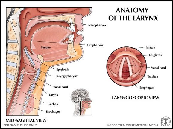
The study of humor and laughter, and its psychological and physiological effects on the human body, is called gelotology. Neurophysiology, the study of the nervous system function, indicates that laughter is linked with the activation of the ventromedial prefontal cortex, which produces endorphins (hormones) and are associated with pure emotion regulation. Endorphins triggered by laughter not only relieve the pain of accident or illness, but they can actually enhance the healing process by helping us develop greater optimism and joy.
Picture:
http://www.trialsightmedia.com/exhibit_store/index.phpmain_page=product_info&products_id=34
Picture:
http://www.trialsightmedia.com/exhibit_store/index.phpmain_page=product_info&products_id=34
Vetromedial Prefrontal Cortex
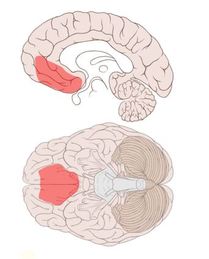
The vertormedial prefrontal cortex is located in the frontal lobe of the brain and is in charge of processing fear, risk, and decision making. Neural networks in the ventromedial prefrontal cortex quickly develop during adolescence and young adulthood, supporting emotion regulation through the amyglada. The left side of the prefrontal cortex is highly active during guessing tasks, while the right side is associated with regulating reasoning and responses.
Picture:http://www.trialsightmedia.com/exhibit_store/index.php?main_page=product_info&products_id=34
The Limbic System
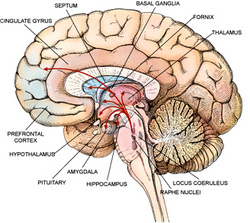
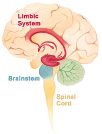
The Structures in the Limbic System Involved in Laughter:
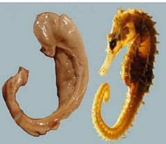
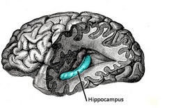
The Hippocampu s is a major component of the brains of humans and other mammals. It is located inside the medial temporal lobe. It plays important roles in the putting together of information from short-term memory to long-term memory and spatial navigation. It sends memories out to the appropriate part of the cerebral hemisphere for long-term storage and retrieves them when necessary.
Picture:http://en.wikipedia.org/wiki/File:Hippocampus_and_seahorse.JPG
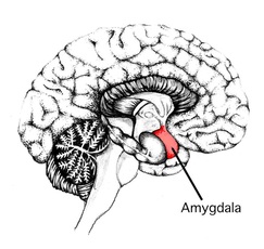
The Amygdala is responsible for determining what memories are stored and where the memories are stored in the brain. It is believed that this determination is based on how big of an emotional impact the event has on the person. It is also responsible for arousal, autonomic responses associated with fear, emotional responses, and hormonal secretion.
Picture: http://www.liminalmusic.org/index.php?/files/amygdala1/2/
Sources:
http://www.msnbc.msn.com/id/31802854/ns/health-behavior/t/ha-laughter-comes-two-types-study-says/
http://biology.about.com/od/anatomy/a/aa042205a.htm
http://webspace.ship.edu/cgboer/limbicsystem.html
http://men.webmd.com/features/why-we-laugh?page=2
http://biology.about.com/od/anatomy/p/Amygdala.htm
http://www.biopsychiatry.com/amygdala.htm
http://www.memorylossonline.com/glossary/hippocampus.html
http://faculty.washington.edu/chudler/hippo.html
http://biology.about.com/od/anatomy/p/hippocampus.htm
http://www.associatedcontent.com/article/5760396/the_mystery_of_laughter_the_brain_plus.html?cat=5
